Ecg Changes In Mi : Figure 16. ST segment elevations. - ECG learning / Serial, morphologic, typical qrst changes with time after transmural mi are depicted.
Ecg Changes In Mi : Figure 16. ST segment elevations. - ECG learning / Serial, morphologic, typical qrst changes with time after transmural mi are depicted.. The four main changes all doctors and medical students should know inside out that occur in a stemi.there are different changes in posterior mi, nstemis and. Recognition of the ecg/ekg changes of hyperkalemia can save lives. Reversibility (ischemia vs infarction) 6 ischemia: Think of it as a stream and someone has put a big rock in the middle. No obviously evident ecg changes (there may be some transient changes), negative troponin, often a history suggestive of acs.
Unstable angina is significant due to the high risk (50%) of mi in the subsequent 30 days. Download scientific diagram | evolution of ecg changes in mi. Arrhythmias other than vt including multifocal pvcs, triplets. M.i denotes cellular damage due to prolonged ischaemia.sudden cardiac death is a frequent presenting feature of mi. What you need to do at emergency room?
Diagnostic ECG—The 12-Lead (Clinical Essentials ... from what-when-how.com There are five basic acute mi ecg patterns you will encounter. Both produce the same spectrum of ecg changes and symptoms and are managed identically in the emergency department. Download scientific diagram | evolution of ecg changes in mi. Tall, positive, hyper acute t waves in the ischemic vascular territory. Also, it can distinguish clinically different types of myocardial infarction. M.i denotes cellular damage due to prolonged ischaemia.sudden cardiac death is a frequent presenting feature of 5. 1.the earliest changes are : They are grouped together because.
So detection of elevated serum cardiac enzymes is more important than ecg changes.
3 ecg on diagnosis of mi cornerstone in diagnosis of acute and chronic ihd factors: For example, an active change in leads v3 and v4, suggesting a problem in the left anterior descending artery and affecting the ventricular septum, would be expected to cause reciprocal changes in leads ii, iii, and avf. Nonspecific ecg changes in acute stroke has therefore posed much diagnostic and. 2.this is followed by elevation of. Scoring of mi for mortality in first 14 days. So, this pattern is what we call a normal ecg. It is a graph of voltage versus time of the electrical activity of the heart using electrodes. 18.04.2017 · ecg changes in mi 1. The ecg changes are often critical in the diagnosis of acute mi and guiding therapy. Study flashcards on ecg changes in mi at cram.com. M.i denotes cellular damage due to prolonged ischaemia.sudden cardiac death is a frequent presenting feature of 5. What you need to do at emergency room? Cardiac markers in acute mi.
For example, an active change in leads v3 and v4, suggesting a problem in the left anterior descending artery and affecting the ventricular septum, would be expected to cause reciprocal changes in leads ii, iii, and avf. When the paradigm changed to stemi vs nstemi, posterior mis were still missed because they didn't produce st elevation on the 12 lead. Unstable angina is significant due to the high risk (50%) of mi in the subsequent 30 days. Cram.com makes it easy to get the grade you want! It is a graph of voltage versus time of the electrical activity of the heart using electrodes.
ECG - MI. Acute Coronary Syndromes Unstable Angina ... from cf2.ppt-online.org When the paradigm changed to stemi vs nstemi, posterior mis were still missed because they didn't produce st elevation on the 12 lead. Study flashcards on ecg changes in mi at cram.com. Scoring of mi for mortality in first 14 days. No obviously evident ecg changes (there may be some transient changes), negative troponin, often a history suggestive of acs. Nonspecific ecg changes in acute stroke has therefore posed much diagnostic and. They are grouped together because. 3 ecg on diagnosis of mi cornerstone in diagnosis of acute and chronic ihd factors: Ecg changes during myocardial infarction (mi).
Tall, positive, hyper acute t waves in the ischemic vascular territory.
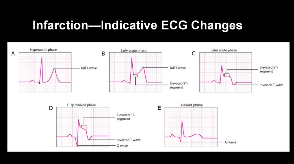
So detection of elevated serum cardiac enzymes is more important than ecg changes. Jesse maclaren reviews posterior mi ecg interpretation on em cases' ecg cases blog. Not to be confused with stable angina. 3 ecg on diagnosis of mi cornerstone in diagnosis of acute and chronic ihd factors: Cardiac markers in acute mi. Arrhythmias other than vt including multifocal pvcs, triplets. Serial, morphologic, typical qrst changes with time after transmural mi are depicted. There is a series of ecg changes reflect the evolution of the infarction (the figure below). Cram.com makes it easy to get the grade you want! When the paradigm changed to stemi vs nstemi, posterior mis were still missed because they didn't produce st elevation on the 12 lead. M.i denotes cellular damage due to prolonged ischaemia.sudden cardiac death is a frequent presenting feature of 5. So, this pattern is what we call a normal ecg. Download scientific diagram | evolution of ecg changes in mi.
Not to be confused with stable angina. Electrocardiography is the process of producing an electrocardiogram (ecg or ekg). 2.this is followed by elevation of. Severe ischemia results in ecg changes within minutes. The ecg changes associated with acute pulmonary embolism may be seen in any condition that causes acute pulmonary hypertension, including hypoxia causing pulmonary hypoxic vasoconstriction.
PPT - ECG Changes in Acute Myocardial Infarction ... from image.slideserve.com It is a graph of voltage versus time of the electrical activity of the heart using electrodes. Ecg changes in ischemia are usually of t wave inversion, st depression or both together. Ecg changes and indicators mi. Think of it as a stream and someone has put a big rock in the middle. Jesse maclaren reviews posterior mi ecg interpretation on em cases' ecg cases blog. The ecg changes in this phase are 1.tall,symmetrical,peaked and widened t waves 2.slope elevation of st segment 3.increased amplitude of r wave/changes in. M.i denotes cellular damage due to prolonged ischaemia.sudden cardiac death is a frequent presenting feature of mi. Both produce the same spectrum of ecg changes and symptoms and are managed identically in the emergency department.
We look for changes in this pattern.
There is a series of ecg changes reflect the evolution of the infarction (the figure below). Recognition of the ecg/ekg changes of hyperkalemia can save lives. They are grouped together because. Electrocardiography in suspected myocardial infarction has the main purpose of detecting ischemia or acute coronary injury in emergency department populations coming for symptoms of myocardial infarction (mi). It is a graph of voltage versus time of the electrical activity of the heart using electrodes. M.i denotes cellular damage due to prolonged ischaemia.sudden cardiac death is a frequent presenting feature of 5. 18.04.2017 · ecg changes in mi 1. When the paradigm changed to stemi vs nstemi, posterior mis were still missed because they didn't produce st elevation on the 12 lead. M.i denotes cellular damage due to prolonged ischaemia.sudden cardiac death is a frequent presenting feature of mi. Jesse maclaren reviews posterior mi ecg interpretation on em cases' ecg cases blog. Download scientific diagram | evolution of ecg changes in mi. What to look for in ecg? Unstable angina is significant due to the high risk (50%) of mi in the subsequent 30 days.
Belum ada Komentar untuk "Ecg Changes In Mi : Figure 16. ST segment elevations. - ECG learning / Serial, morphologic, typical qrst changes with time after transmural mi are depicted."


Belum ada Komentar untuk "Ecg Changes In Mi : Figure 16. ST segment elevations. - ECG learning / Serial, morphologic, typical qrst changes with time after transmural mi are depicted."
Posting Komentar